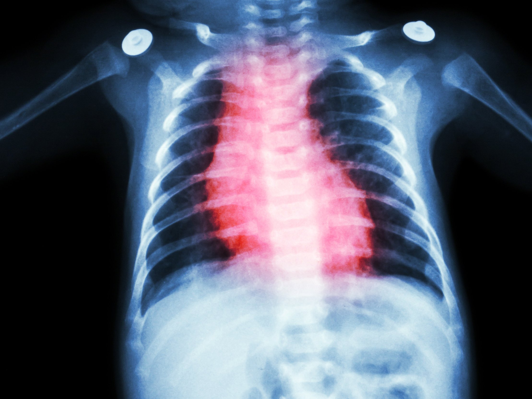
A Broken Heart: Hypoplastic Left Heart Syndrome
Despite the advances in congenital heart disease management, hypoplastic left heart syndrome (HLHS) remains the most difficult to palliate long-term and overall survival of these patients is approximately 50% (Fraser, 2015).
Hypoplastic left heart syndrome is defined as a congenital cardiac malformation where the left ventricle, aortic valve, and aortic root are inadequate to perfuse systemic circulation. In other words, the infant has little chance of survival without staged, complicated surgeries. Even with these surgeries, the patient may need a heart transplant to survive (Rychick, 2014; Fraser, 2015).
In the past a diagnosis of HLHS was a death sentence, infants were lovingly taken home to die. Nowadays, with the advent of prenatal echocardiography, prenatal diagnosis is available. This allows for detailed evaluation of structure and function during gestation. Diagnosis in-utero allows the parents and family to adjust and plan for the care of the infant with HLHS. The medical team can plan for the immediate stabilization of the infant in preparation for the first surgical intervention.
HLHS Anatomy:
In-utero, the left side of the heart fails to develop normally resulting in a combination of defects and severity of defects. HLHS defects include: left ventricular hypoplasia, mitral and aortic valve atresia or stenosis, hypoplastic aorta, and aortic arch. Survival is dependent on the severity of the defects; the left heart can be either poorly developed or non-existent (Rychick, 2014; Fraser, 2015).
In-utero, circulation is maintained via the ductus arteriosus which shunts blood into the systemic circulation.
HLHS Physiology:
It is important to understand the differences in ventricular function. The right ventricle’s main function is to eject volume into the lungs against pulmonary vascular resistance (less than 250 dynes sec/cm5); while the left ventricle ejects volume against systemic vascular resistance (800-1200 dynes sec/cm5). This difference in pressure results in increased muscle mass in the left ventricle. In severe HLHS, the right ventricle must become the systemic pump. This change in function may lead to progressive right ventricular failure or dysfunction (Bowden & Greenberg, 2014; Rychick, 2014; Fraser, 2015).
HLHS Management:
After birth, depending on the severity of the HLHS defects, the ductus arteriosus must remain patent. Intravenous prostaglandins to maintain the patency of the ductus arteriosus and to decrease pulmonary hypertension, and other medications such as vasoactive agents may be needed to stabilize the infant in preparation for the first of three surgeries. Intubation and mechanical ventilation with nitrogen and oxygen combinations to lower the oxygen concentration or PaO2 to balance pulmonary and systemic vascular resistance (Rychick, 2014; Fraser, 2015).
Surgical Palliation:
Surgical palliation begins at about 2 weeks of age with Stage I: the Norwood Procedure. Stage II is the bidirectional Glenn Shunt; usually completed at age 4-months, and Stage III is the Fontan procedure; usually performed around age 2-4 years (Bowden & Greenberg, 2014; Rychick, 2014; Fraser, 2015).
Stage I: Norwood Procedure:
To provide viable systemic circulation, the atrial septum must be opened to allow the mixing of the oxygenated blood and unoxygenated blood; bypassing the left ventricle. Next, the main pulmonary artery is connected to the aorta allowing the mixed oxygenated blood to be delivered to the systemic circulation via the pulmonary valve. Because of these changes, a shunt must be provided to allow blood flow to the pulmonary circulation. This procedure is called a Blalock-Taussig shunt; a connection from the subclavian artery to the pulmonary artery is accomplished by the addition of a Gore-Tex conduit.
The blood flow is now directed in this manner:
Unoxygenated blood returns to the right atria where it mixes with the oxygenated blood from the left atria. This mixed oxygenated blood flows into the right ventricle via the tricuspid valve and is pumped to the systemic circulation; supplying the tissues with needed nutrients and oxygen. Additionally, at the level of the subclavian artery, blood is shunted into the pulmonary circulation to become oxygenated in the lungs and returned to the left atria. This series of shunts allow the infant to grow while strengthening the right ventricle and allowing pulmonary vascular resistance to decrease. When the pulmonary vascular resistance is lowered, Stage II palliation may occur (Bowden & Greenberg, 2014; Rychick, 2014 & Fraser, 2015).
The infant born with hypoplastic left heart syndrome will require several surgical procedures to provide adequate systemic oxygenation. In this article we looked at the Norwood procedure, the first stage in the multistage approach to palliate HLHS. In the second article, we will discuss stage two: Norwood Stage II also known as a bi-directional Glen or Hemi-Fontan.
References
Bowden, V. & Greenberg, C. (2014). The child with altered cardiovascular status in Children and their families: the continuum of nursing care, third edition, pp. 645-646. Fraser, C. (2015). The journey toward improved hypoplastic left heart syndrome outcomes continues-another small step. The Journal of Thoracic and Cardiovascular Surgery; 149:6, pp.1487. Rychick, J. (2014). Hypoplastic left heart syndrome: Can we change the rules of the game?


